
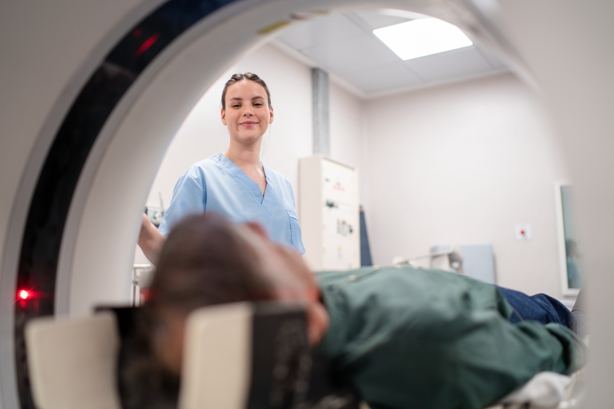
The Emergency Department at UAB St. Vincent’s East recently installed an advanced CT scanner that produces more detailed images of the body with lower radiation doses.
Computed tomography (CT) scanners combine X-rays with computer technology to create 3D images of organs, bones, and tissue inside the body. A radioactive contrast material is first injected into the patient, then the scanner captures images as this material moves through the body.
CT scans are widely used in health care to locate and evaluate injuries, tumors, blood clots, and many other conditions. Features of this system include:
- Cameras connected to the machine enable better positioning of the patient and closer monitoring during the scan.
- The scanner’s premium X-ray system allows for less contrast material and lower radiation dosages to be used.
- A set of colored lights indicates to patients when they should hold their breath, which results in more detailed images.
“Patients are our number one priority,” said Mark Hyatt, director of Radiology and Imaging at UAB St. Vincent’s East. “We carefully chose a CT system that would provide the best possible experience for our patients and give our physicians the information they need to deliver the best possible care.”